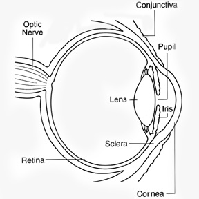
The cornea is clear and seems to lack substance, it is actually a highly organized group of cells and proteins. Unlike most tissues in the body, the cornea contains no blood vessels to nourish or protect it against infection. Instead, the cornea receives its nourishment from the tears and aqueous humor that fills the chamber behind it. The cornea must remain transparent to refract light properly, and the presence of even the tiniest blood vessels can interfere with this process. To see well, all layers of the cornea must be free of any cloudy or opaque areas.
Q. How does the eye work?
A. Anything you see in image that enters your eye in the form of light the different parts of your eye collect this and send a message to you brain. Enabling you it sees for perfect vision all the parts of your eye need to work properly.
Q. What is a Cornea?

A. The cornea is the eye's outermost layer. It is the clear, dome-shaped surface that covers the front of the eye.
Q. What is the function of Cornea?
A. Because the cornea is as smooth and clear as glass but is strong and durable, it helps the eye in two ways: It helps to shield the rest of the eye from germs, dust, and other harmful matter. The cornea shares this protective task with the eyelids, the eye socket, tears, and the sclera, or white part of the eye. The cornea acts as the eye's outermost lens. It functions like a window that controls and focuses the entry of light into the eye. The cornea contributes between 65-75 percent of the eye's total focusing power. When light strikes the cornea, it bends--or refracts--the incoming light onto the lens. The lens further refocuses that light onto the retina, a layer of light sensing cells lining the back of the eye that starts the translation of light into vision. For you to see clearly, light rays must be focused by the cornea and lens to fall precisely on the retina. The retina converts the light rays into impulses that are sent through the optic nerve to the brain, which interprets them as images. The refractive process is similar to the way a camera takes a picture. The cornea and lens in the eye act as the camera lens. The retina is similar to the film. If the image is not focused properly, the film (or retina) receives a blurry image. The cornea also serves as a filter, screening out some of the most damaging ultraviolet (UV) wavelengths in sunlight. Without this protection, the lens and the retina would be highly susceptible to injury from UV radiation.
Q. How does the Cornea respond to injury?
A. The cornea copes very well with minor injuries or abrasions. If the highly sensitive cornea is scratched, healthy cells slide over quickly and patch the injury before infection occurs and vision is affected. If the scratch penetrates the cornea more deeply, however, the healing process will take longer, at times resulting in greater pain, blurred vision, tearing, redness, and extreme sensitivity to light. These symptoms require professional treatment. Deeper scratches can also cause corneal scarring, resulting in a haze on the cornea that can greatly impair vision. In this case, a corneal transplant may be needed.
Some diseases and disorders affecting the Cornea:
Allergies Conjunctivitis (Pink Eye) Corneal Infections Dry Eye Fuchs' Dystrophy Herpes Zoster (Shingles) Iridocorneal Endothelial Syndrome Keratoconus Lattice Dystrophy Map-Dot-Fingerprint Dystrophy Ocular Herpes Pterygium Stevens-Johnson Syndrome
Q. What is a Corneal Transplant?
A. A transplant is the replacement of damaged or diseased tissues or organs with healthy tissues or organs In corneal transplant the cloudy or warped cornea is replaced with a healthy cornea if the new cornea heals without problems there con be tremendous improvement in vision. Since the cornea was one of the first parts of the body to be transplanted corneal transplants remain one of the most common and most successful of all transplants.

Q. Who supplies the healthy cornea?
A. The corneal used for transplantation is supplied by an Eye Banks Eye Banks work round the clock to collect. Evaluate and store donated corneas The corneas are collected from human donors within hours of death stringent tests are done to ensure the safety of the person receiving the cornea. The Eye Bank verifies the donor medical history and cause of death and performs blood tests to ensure that the deceased person did not have any contagious disease such as AIDS or hepatitis.
Facts you may like to know
- It is not necessary to find cornea with a matching tissue or blood type.
- The race gender and eye color of the donor are not important.
- A corneal transplant won t change your natural eye color.
- The cornea heals slowly and improvement in vision may take a year or more.
- It is difficult to shape the new cornea perfectly so astigmatism (a condition where the cornea has an irregular shape making images seems blurred to distort) is common after a corneal transplant However. This can be corrected with glasses.
Scheduling a transplant
If you are advised to undergo a corneal transplant your ophthalmologist will tell about the procedure. The transplant will be scheduled according the condition of you eye and availability of a donor cornea sometimes shortage of donor corneas may delay surgery
If both your eyes need new corneas the second transplants will not be performed unit the first eye has stabilized which may bake up to a year.
Preparing for surgery
If you are taking any other medication ask your ophthalmologist you should continue you will probably be asked not to eat or drink anything for several hours before the surgery.
Usually local anesthesia used so you will be awake but feel no pain intravenous medications will help your relax the nerves in your will be completely so numbed so you will not be able see or move your eye. Sometimes the doctor may sue general anesthesia.
The transplant procedure
For the transplant doctors use an operating microscope and very delicate instruments once the old cornea is removed the now cornea is stitched into place the sutures or stitches are barely visible and are not painful although you may feel some irritation or a scratching sensation for a few days.
If necessary other procedure may be performed at the some time as your transplant your ophthalmologists will advise you about them.
Potential risk
As with other surgical a corneal transplant some risks and most of them con be treated some possible complications are.
- Eye infections
- Failure of the donor cornea to function normally
- Rejection of the donor cornea by your body
- Cataract (clouding of the eyes lens)
- Glaucoma (build -up of fluid leading to increased pressure in the eye)
- Bleeding from the iris
- Swelling or detachment of the retina
Rejection the danger signals
Rejection of a transplanted cornea can occur any time but is more likely to happen in the fist year after surgery reduces the chance of success of any repeat corneal transplantation however rejection can be prevented by timely diagnosis and appropriate management. Watch out these danger signals.
- Redness
- Sensitivity to light
- Vision loss
- Pain
The acronym RSVP can help you remember symptoms if you notice any of these in your operated eye. However minor they may seem and regardless of the time of day, contact us immediately. If this is not possible, visit an ophthalmologist, preferably a cornea specialist.
Care after surgery
- Do not lift heavy things.
- Do not bend so that you head is lower then you waist.
- Avoid sleeping on the operated side.
- No sexual intercourse until permitted by the doctor.
- Do not rub the operated eye.
- Avoid any vigorous activity.
- Avoid alcoholic beverages.
- Watch television only for short periods.
You can bathe carefully from below the neck and shave, but do not let the operated eye get wet for at least 15 days. You may gently clear the eyelids with a piece of boiled in water or a sterilized tissue. Do not wet the eyeball. You should wear an eye patch at night, the doctor will advise you when to discontinue using it during the day. Always wear protective glasses or an eye shield to avoid accidental injury.
Medication and follow-up
At the time of discharge our patient counselor will advise you about medication and follow-up visits. Please follow the instructions regarding medication; please adhere to the follow up appointment date.
If you have any concerns or questions, ask the doctor when you an examination, if you feel you cannot wait, call or email us at our numbers given below.
If this is an emergency at right, during a weekend, or on a holiday, come for emergency care to the institute.
Always mention the patient ID number, name and doctor's name in all communications.
Concerned Doctors in CLGEI:


